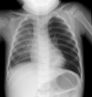
Uses Of Plain Child Dental X Ray
Unquestionably, various innovations like Computed
Tomography (CT), Magnetic Resource Imaging (MRI) and Ultrasound Imaging (UI)
have surfaced throughout the years in the medicinal business. Be that as it
may, plain x-beams hold their significance till date with regards to diagnosing
various types of sicknesses. Plain x-beams or radiography include utilization
of a x-beam generator to create a x-beam pillar which goes through the required
protest like the body part that should be inspected. The question or material
that the pillar infiltrates assimilates the same with the sums fluctuating
according to the thickness and structure of the protest. The x-beams that don't
get ingested get recorded on a film.
For the most part, it's the bones of the patient
that effectively assimilate the x-beams to seem white on the picture, while
delicate tissues like muscle fiber retain a lesser measure of x-beams and in
this way, seem dim or grayish. It is this motivation behind why the highly
contrasting complexity is for the most part observed on x-beam pictures.
Additionally, this is the motivation behind why traditional or plain x-beams
are esteemed as proper for bone or tissue checks, be it investigation of a bone
break or ct scan radiation risk imagegently dental imaging.
Radiography or plain x-beam is likewise used to
inspect the stomach area organs like bladder or liver. Further, it encourages
lead chest radiography to recognize any lung illnesses, including lung
malignancy and pneumonia. Mammography is another procedure that identifies
bosom tumor at a beginning period. Keeping in mind the end goal to distinguish
sicknesses identified with digestive system or stomach or urinary tract, x-beam
fluoroscopy is brought into child
dental x ray utilization
.
Medicinal x-beam pictures were prior appeared on
photographic film. The same required a bit of preparing before the picture
could be seen. Notwithstanding, advanced x-beams are dissimilar to the same and
utilize an electronic locator. A PC is utilized to process the picture which
can be put away carefully. Likewise, the specialist can see the same right
away.
Utilizing plain x-beams, specialists can get
high-determination, quick pictures at reasonable expenses. A plain x-beam test
does not take over 15 minutes. What the radiographer should simply pick the
sort of x-beams for utilize and measure of x-beams according to the extent of
the patient, the body part to be analyzed and the picture differentiate. The
reason being the development of the body part makes the picture obscured. The
picture delivered gets put away on radiograph and a radiologist investigates
the same.
In any case, the x-beam picture creation includes
use of ionizing radiation which is cancer-causing enough to make some sort of
harm patient's wellbeing if presented to it. In any case, specialists say that
the advantages of radiography are sufficiently effective to dominate the slight
dangers associated with the methodology.
A x-beam test is typically viewed as more unsafe
with regards to directing the same on a pregnant lady. Since, the fetus is
exceedingly delicate to the radiations the lady is presented to. This may bring
about some sort of variation from the norm or other medical issue in the
unborn.


Comments
Post a Comment